Sunday, November 18, 2012

Copper To The Rescue?

Jane M Flinn
Dr. Jane Flinn

        Our lecturer this week was Dr. Jane M. Flinn, director of undergraduate neuroscience program at George Mason University. Dr. Flinn's current research focuses on the role of 
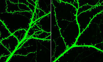
metals in normal memory and in Alzheimer's disease (AD). The brain of those with AD contains plaques and tangles. The plaques contain amyloid, a protein which is aggregated by zinc, but which also binds copper and iron.

        Recently, she observed elevated levels of zinc, iron and copper in the plaques found in brains of people with AD.  The transgenic mice she experimented with carried an APP mutation so unlike most mice, they developed plaques. By administering different levels of metals in their drinking water, her team's results showed "both zinc and iron significantly impaired spatial memory in mice modeling early onset AD but copper partially remediated the zinc effect." Additionally, increased zinc diminishes the ability to learn that a stimulus is no longer fearful in normal mice and rats. This effect can be a model of post-traumatic stress disorder (PTSD). Small amounts of copper have been shown to partially alleviate these symptoms. Perhaps learning impairment could be a result of copper deficiency?

A soldier affected by PTSD
        Previous research have demonstrated chronic stress decreases zinc in the blood while increases it in the brain. This suggests zinc might be redistributed from the blood to the brain. Results from other previous research showed excess zinc both pre and post-natally demonstrated impairments in fear extinction thus zinc may be a mediator between stress and the inability to extinguish fear.

        Dr. Flinn conducted the experiment, The Effects of Chronic Unpredictable Stress (CUS) on the Ability to Extinguish Fear: Zinc as a Mediator, where they collected data from mice given zinc and measured impairments in fear extinction. The subjects were 31 Sprague-Dawley rats bred both pre and post-natally on either water enhanced with zinc (10mg/kg ZnCO3) or tap water. The four groups were: tap water + stress (control), tap water + stress, zinc + no stress, zinc + stress. A 21 day randomized chronic unpredictable stress paradigm was administered. 10 days post stress, cued fear conditioning was conducted. Day 1: training = 3 tone + shock pairings. Day 2: extinction = 18 tones and no shock. Day 3: recall = 18 tones and no shock.
Representative metal output images. Images show iron
(Fe, top left), calcium (Ca, top right),
zinc (Zn, bottom left), and potassium (K, bottom right).
(White color= greatest concentration).

        Dr. Flinn and her team concluded the zinc group took longer to learn fear extinction as well as memory deficits with impairment of recall. CUS rats showed less freezing when anticipating shocks compared to control group. This concludes a down regulation of the HPA axis even 10 days post termination of stress.

         The work of Dr. Flinn will guide future directions of these types of research. If increased Zinc in the diet can cause deficiency, are the deficits in the fear conditioning due to a copper deficiency? Thus far we think the deficits in AD are due to a copper deficiency, continuing work on developing mouse model of late onset AD would confirm or reject this hypothesis. It is suggested that AD is caused by an inflammation problem. Dr. Flinn mentioned an incident where there were two identical twins and one of them took aspirin regularly. The other developed AD 10 years sooner than the twin on the aspirin regimen. This, again, raises new question that will hopefully be answered with further research.


Reference

The Effects of Chronic Unpredictable Stress (CUS) on the Ability to Extinguish Fear: Zinc as a Mediator
Knaack, G.L., McDonald, C.G., & Flinn, J.M. Dept. Psychology, George Mason Univ., Fairfax, VA, USA

Sunday, November 4, 2012

Alpha 7/GPRIN1 To Infinity And Beyond

         Our brain's wiring makes us who we are. Without a doubt, this process affects all aspects of our lives. The brain lays the foundation of our neural pathways during development. What structures are responsible for this incredible task? What symptoms would occur if they are damaged? What can we discover from researching the processes involved?


Jacob Nordman, Ph.D
         On November 1st, 2012, George Mason University's Neuroscience Ph.D. Candidate from Dr. Kabbani's lab, Jacob Nordman, presented his research regarding alpha 7 nAChR and Gprin1 to our NEUR410 course. The lecture focussed on alpha 7 and Gprin1 interaction which regulates axon growth and growth cone dynamics on hippocampal neurons. The objective of Kabbani's lab and Nordman's research is to study the proteome and their relation to dopamine and nicotinic acetylcholine receptors in the nervous system. Neural activity involving these receptors play a vital role in complex brain functions including cognition, attention, and memory. As a result, studying them should further our understanding of drug development for the treatment of several human brain disorders.

A) Ionotropic receptor. B) Metabotropic receptor
          Before describing Nordman's research, a few background concepts should be explained. Ionotropic and metabotropic receptors vary in speed and duration of their effects. Ionotropic binds a neurotransmitter, opens the channel, allows an immediate flow of ions, and induces an EPSP or IPSP. Metabotropic binds a NT and allows a cascade of secondary messenger systems to occur. This may include but is not limited to: 1) opening another channel via an internal binding site, 2) increasing or decreasing transcription, and 3) protein modifications, including phosphorylation. Furthermore, scaffold proteins act as crucial regulators of these many signaling pathways. Their functions include opening new signaling cascades, act as new drug targets, novel net plasticity mechanisms, provide greater inter-connectivity between neurons, and even more functions we have yet to discover.
Growth cone structure

          The specific focus of Nordman's research is on the growth cone. It is a complex structure composed of three layers which guides axon development. The seven states it can be found in are initiation, formation, guidance, branching, turning, arrest, and retraction. Each growth cone state performs the function their name implies.

          Mr. Nordman and his team have found that alpha 7 nAChR (nicotinic acetylcholine receptor) are enriched within growth cones. He research also focuses on a newly discovered cytoskeletal regulator termed G protein regulated inducer of neurite outgrowth 1 (GPRIN1). GPRIN1, also enriched in growth cones, scaffolds nAChRs within neurons. This brings us to his current study which investigates how alpha 7 nAChR regulates growth cone dynamics and axon targeting in the cortex and hippocampus during early brain development. The techniques being used include subcellular fractionation to isolate growth cones from newborn pups for proteomic analysis. They observed alpha 7 nAChR/GPC in the hippocampus under a microscope by highlighting it with anibodies fluorescent, a yellow signal.


          When in development were these proteins present? Nordman responded, the highest expression was during the guidance period. It was most abundant in the soma/GC which makes sense because that is where we expect the highest demand. How can we prove alpha 7/GPC interactions were present in the GC? Nordman stated neuron2a were neuron-like cells. He used transfection, planting DNA into a cell to produce protein; followed by immunoprecipation, removing many subunits of cells to focus on relevant network he was measuring. By eliminating GP1 from scaffold to limit interaction with GP1 and alpha 7, the weakened link suggested they are in fact connected. To confirm active apha nAChR are present in the growth cone, Nordman injected calcium sensors in the tissue grown in the petri dish. Using PNU282987, an alpha 7 activator, a green fluorescent was present which suggested alpha 7 was, in fact, in these cells.
          While further investigating their interactions, Nordman stated GPRIN1 is the master switch for alpha 7 signaling. During a 12 hour test, he eliminated the expression of GPRIN1 which resulted in shorter and less branching of neurons. This suggested GPRIN1 and alpha are complementary and both are vital in growth cone function. To mediate alpha 7 growth, EB3 (end binding) comets, a microtubule capping protein, was used. Using time lapse to give us real time changes of growth, EB3 moved at approximately 3-5 microns every 10 seconds.

          Mr. Nordman concluded that in "active state", filopodia projects in all directions. It commits to a direction through microtubules invasion or collapse filopodia. Alpha 7 activation inhibits G proteins mediated pathways involved in growth. The sum effect is microtubule capping and growth cone collapse.

Normal compared to Schizophrenic neuron
Fewer pathways cause symptoms of schizophrenia.
This image shows neuron pathway (green strands)
comparison between a healthy mouse (left)
and one bredto express schizophrenia (right).
          Based on Nordman's findings, could it be possible to reverse this phenotype by inhibiting alpha 7 and prevent neuronal damage and promote axonal growth? He emphasized the incredibly complicated process of regenerating axons but was optimistic that it is possible. Will it have any effects on multiple sclerosis since it mainly affects myelin sheath? Perhaps an alpha 7 antagonist could be the cause of promoting axons regeneration that will effectively treat or cure schizophrenia, alzheimer's or spinal cord injuries?


Reference: http://krasnow.gmu.edu/kabbani/research-2/