Sunday, December 9, 2012

Blasting Neuroblastoma


Alex J. Carlisle
Dr. Alex Carlisle
        Dr. Alex Carlisle is a neuroscientist in the Department of Neurosciences at Inova Fairfax Hospital and head of the Laboratory of Neuro-Oncology at Krasnow. He is a cancer biologist who uses molecular-based approaches to identify and functionally characterize molecules involved in the progression of various cancers. On December 6th, 2012, he presented a power point presentation to our NEUR410 course discussing his current research: Biological and Clinical Association of CXCR4 with the Development of Peripheral Neuroblastic Tumors (pNTs).
        
        Neuroblastoma is a disease where malignant (cancerous) tumors start to develop in the sympathetic nervous system and may spread to all other parts of the body. It mainly affects infants and children. The cause of this tumor is currently unknown but Dr. Carlisle explained Phox2B, a gene found in the development of sympathoadrenal cell lineage, may be an indicator of this disease.
        
Anatomy of the female urinary system; drawing shows a front view of the right and left kidneys, the ureters,  urethra, and bladder filled with urine. The inside of the left kidney shows the renal pelvis. An inset shows the renal tubules and urine. The spine, adrenal glands, and uterus are also shown.
Neuroblastoma is a disease
in which malignant (cancer) cells
form in nerve tissue of the
adrenal gland, neck, chest,
or spinal cord.
        Neuroblastoma is the number one cancer found in infants, accounting for approximately 10% of all pediatric cancers and 15% of cancer related deaths in children. Early detection is vital but too often when patients are first diagnosed, it has already spread. Children who are diagnosed at 1 year old or less have the best possible outcome. The severity of their diagnosis ranges from Stage I (isolated cancer) to Stage IV (metastasis). Studies have shown on rare occasion, stage IV neuroblastoma sometimes undergoes spontaneous regression without therapeutic intervention. “We don’t know why this happens”, said Carlisle. Treatments include surgery, chemotherapy, and/or radiation. Statistics show more powerful biomarkers and improved therapies are urgently needed to diagnose neuroblastoma. What role does Carlisle’s research play in all this?

        The Carlisle laboratory studies how molecules in tumor cells from patients interact to promote the aggressive behavior of neuroblastoma. This approach allows Carlisle to identify biomarkers which may aid in improving diagnosis and prognosis. The heterogeneity of this disease makes is very difficult to study but Carlisle has identified a chemokine receptor, CXCR4, whose expression is clinically correlated with advanced stages of neuroblastoma.

Types of Neuroblastoma
        The biological roles for CXCR4 include inflammation (lymphocyte homing and recruitment into inflammatory sites), neuronal development (NPC migration during embryogenesis), metastasis, HIV infection (co-receptor for HIV binding and fusion; CD4+ cells), and cancer progression. Would knocking out CXCR4 solve the problem? No, CXCR4 is necessary for normal neuronal formation and development of an organism. A genetic knockout would yield growth malformation of the dorsal root ganglia which would kill the organism.

CT image of neuroblastoma tumor
        A variety of clinical and biological parameters are used for risk-assessment and outcome prediction of neuroblastoma. Carlisle stressed the importance of identifying the stage of the cancer which greatly helps initiate proper treatments. His research showed CXCR4 protein expression was least in ganglioneuroma (benign tumor) and progressively increased as the stages worsened with the highest expression in Stage IV neuroblastoma. This suggests CXCR4 is highly involved in signaling cancer development to metastasis.

        In an attempt to draw correlations with transcription factors, Carlisle found little evidence to show there was any significance. Do CXCR4 and MYCN interact? MYCN is a gene associated with a variety of tumors, most notably neuroblastomas. I would assume an amplification of this gene would present with higher levels of CXCR4 and neuroblastomas but Carlisle’s data showed no correlation between the two.

        Despite numerous roadblocks, Carlisle was able to find a successful treatment for these cancer cells while evaluating CXCR4-mediated signaling pathways associated with neuroblastoma progression. Plerixafor blocks CXCR4 (receptors for only CXCL12) and as a result, inhibits growth and migration of cancer cell and tumor growth. It is presumed this is accomplished by preventing macrophages from being recruited to tumors.

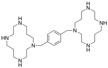
Chemical structure of Plerixafor
        It seems there is a lot of potential in the research Carlisle and his team are doing. Their research on receptor-mediated efficacy responses to the CXCR4-selective antagonist, Plerixafor, suggested this drug treatment is effective against the number cancer found in infants. While studying neuroblastoma maturation, they have also discovered a greater number of Schwann cells yielded to less malignant tumors and better outcome. This opens a new door to study neuroblastoma’s relationship with Schwann cells. Dr. Carlisle has encourages students interested in conducting translational research in the area of Neuro-oncology to contact his laboratory.


Sunday, November 18, 2012

Copper To The Rescue?

Jane M Flinn
Dr. Jane Flinn

        Our lecturer this week was Dr. Jane M. Flinn, director of undergraduate neuroscience program at George Mason University. Dr. Flinn's current research focuses on the role of metals in normal memory and in Alzheimer's disease (AD). The brain of those with AD contains plaques and tangles. The plaques contain amyloid, a protein which is aggregated by zinc, but which also binds copper and iron.

        Recently, she observed elevated levels of zinc, iron and copper in the plaques found in brains of people with AD.  The transgenic mice she experimented with carried an APP mutation so unlike most mice, they developed plaques. By administering different levels of metals in their drinking water, her team's results showed "both zinc and iron significantly impaired spatial memory in mice modeling early onset AD but copper partially remediated the zinc effect." Additionally, increased zinc diminishes the ability to learn that a stimulus is no longer fearful in normal mice and rats. This effect can be a model of post-traumatic stress disorder (PTSD). Small amounts of copper have been shown to partially alleviate these symptoms. Perhaps learning impairment could be a result of copper deficiency?

A soldier affected by PTSD
        Previous research have demonstrated chronic stress decreases zinc in the blood while increases it in the brain. This suggests zinc might be redistributed from the blood to the brain. Results from other previous research showed excess zinc both pre and post-natally demonstrated impairments in fear extinction thus zinc may be a mediator between stress and the inability to extinguish fear.

        Dr. Flinn conducted the experiment, The Effects of Chronic Unpredictable Stress (CUS) on the Ability to Extinguish Fear: Zinc as a Mediator, where they collected data from mice given zinc and measured impairments in fear extinction. The subjects were 31 Sprague-Dawley rats bred both pre and post-natally on either water enhanced with zinc (10mg/kg ZnCO3) or tap water. The four groups were: tap water + stress (control), tap water + stress, zinc + no stress, zinc + stress. A 21 day randomized chronic unpredictable stress paradigm was administered. 10 days post stress, cued fear conditioning was conducted. Day 1: training = 3 tone + shock pairings. Day 2: extinction = 18 tones and no shock. Day 3: recall = 18 tones and no shock.
Representative metal output images. Images show iron
(Fe, top left), calcium (Ca, top right),
zinc (Zn, bottom left), and potassium (K, bottom right).
(White color= greatest concentration).

        Dr. Flinn and her team concluded the zinc group took longer to learn fear extinction as well as memory deficits with impairment of recall. CUS rats showed less freezing when anticipating shocks compared to control group. This concludes a down regulation of the HPA axis even 10 days post termination of stress.

         The work of Dr. Flinn will guide future directions of these types of research. If increased Zinc in the diet can cause deficiency, are the deficits in the fear conditioning due to a copper deficiency? Thus far we think the deficits in AD are due to a copper deficiency, continuing work on developing mouse model of late onset AD would confirm or reject this hypothesis. It is suggested that AD is caused by an inflammation problem. Dr. Flinn mentioned an incident where there were two identical twins and one of them took aspirin regularly. The other developed AD 10 years sooner than the twin on the aspirin regimen. This, again, raises new question that will hopefully be answered with further research.


Reference

The Effects of Chronic Unpredictable Stress (CUS) on the Ability to Extinguish Fear: Zinc as a Mediator
Knaack, G.L., McDonald, C.G., & Flinn, J.M. Dept. Psychology, George Mason Univ., Fairfax, VA, USA

Sunday, November 4, 2012

Alpha 7/GPRIN1 To Infinity And Beyond

         Our brain's wiring makes us who we are. Without a doubt, this process affects all aspects of our lives. The brain lays the foundation of our neural pathways during development. What structures are responsible for this incredible task? What symptoms would occur if they are damaged? What can we discover from researching the processes involved?


Jacob Nordman, Ph.D
         On November 1st, 2012, George Mason University's Neuroscience Ph.D. Candidate from Dr. Kabbani's lab, Jacob Nordman, presented his research regarding alpha 7 nAChR and Gprin1 to our NEUR410 course. The lecture focussed on alpha 7 and Gprin1 interaction which regulates axon growth and growth cone dynamics on hippocampal neurons. The objective of Kabbani's lab and Nordman's research is to study the proteome and their relation to dopamine and nicotinic acetylcholine receptors in the nervous system. Neural activity involving these receptors play a vital role in complex brain functions including cognition, attention, and memory. As a result, studying them should further our understanding of drug development for the treatment of several human brain disorders.

A) Ionotropic receptor. B) Metabotropic receptor
          Before describing Nordman's research, a few background concepts should be explained. Ionotropic and metabotropic receptors vary in speed and duration of their effects. Ionotropic binds a neurotransmitter, opens the channel, allows an immediate flow of ions, and induces an EPSP or IPSP. Metabotropic binds a NT and allows a cascade of secondary messenger systems to occur. This may include but is not limited to: 1) opening another channel via an internal binding site, 2) increasing or decreasing transcription, and 3) protein modifications, including phosphorylation. Furthermore, scaffold proteins act as crucial regulators of these many signaling pathways. Their functions include opening new signaling cascades, act as new drug targets, novel net plasticity mechanisms, provide greater inter-connectivity between neurons, and even more functions we have yet to discover.
Growth cone structure

          The specific focus of Nordman's research is on the growth cone. It is a complex structure composed of three layers which guides axon development. The seven states it can be found in are initiation, formation, guidance, branching, turning, arrest, and retraction. Each growth cone state performs the function their name implies.

          Mr. Nordman and his team have found that alpha 7 nAChR (nicotinic acetylcholine receptor) are enriched within growth cones. He research also focuses on a newly discovered cytoskeletal regulator termed G protein regulated inducer of neurite outgrowth 1 (GPRIN1). GPRIN1, also enriched in growth cones, scaffolds nAChRs within neurons. This brings us to his current study which investigates how alpha 7 nAChR regulates growth cone dynamics and axon targeting in the cortex and hippocampus during early brain development. The techniques being used include subcellular fractionation to isolate growth cones from newborn pups for proteomic analysis. They observed alpha 7 nAChR/GPC in the hippocampus under a microscope by highlighting it with anibodies fluorescent, a yellow signal.


          When in development were these proteins present? Nordman responded, the highest expression was during the guidance period. It was most abundant in the soma/GC which makes sense because that is where we expect the highest demand. How can we prove alpha 7/GPC interactions were present in the GC? Nordman stated neuron2a were neuron-like cells. He used transfection, planting DNA into a cell to produce protein; followed by immunoprecipation, removing many subunits of cells to focus on relevant network he was measuring. By eliminating GP1 from scaffold to limit interaction with GP1 and alpha 7, the weakened link suggested they are in fact connected. To confirm active apha nAChR are present in the growth cone, Nordman injected calcium sensors in the tissue grown in the petri dish. Using PNU282987, an alpha 7 activator, a green fluorescent was present which suggested alpha 7 was, in fact, in these cells.
          While further investigating their interactions, Nordman stated GPRIN1 is the master switch for alpha 7 signaling. During a 12 hour test, he eliminated the expression of GPRIN1 which resulted in shorter and less branching of neurons. This suggested GPRIN1 and alpha are complementary and both are vital in growth cone function. To mediate alpha 7 growth, EB3 (end binding) comets, a microtubule capping protein, was used. Using time lapse to give us real time changes of growth, EB3 moved at approximately 3-5 microns every 10 seconds.

          Mr. Nordman concluded that in "active state", filopodia projects in all directions. It commits to a direction through microtubules invasion or collapse filopodia. Alpha 7 activation inhibits G proteins mediated pathways involved in growth. The sum effect is microtubule capping and growth cone collapse.

Normal compared to Schizophrenic neuron
Fewer pathways cause symptoms of schizophrenia.
This image shows neuron pathway (green strands)
comparison between a healthy mouse (left)
and one bredto express schizophrenia (right).
          Based on Nordman's findings, could it be possible to reverse this phenotype by inhibiting alpha 7 and prevent neuronal damage and promote axonal growth? He emphasized the incredibly complicated process of regenerating axons but was optimistic that it is possible. Will it have any effects on multiple sclerosis since it mainly affects myelin sheath? Perhaps an alpha 7 antagonist could be the cause of promoting axons regeneration that will effectively treat or cure schizophrenia, alzheimer's or spinal cord injuries?


Reference: http://krasnow.gmu.edu/kabbani/research-2/

Monday, October 22, 2012

Translational Research: From Bench To Bedside

Robert Lipsky
Robert Lipsky, Ph. D
          Have you ever taken a medication when you were not feeling well? How do we know it's effective against illnesses? What allowed it to go from the laboratory research setting to your medical cabinet?

          On October 18th, 2012 at George Mason University, my NEUR410 class attended a presentation lecture conducted by Robert H. Lipsky Ph. D, the Director of Translational Research in the Department of Neurosciences from Inova Fairfax Hospital. He discussed various topics relating to his translational research including warfarin (coumadin), clinical depression, and spinal cord injuries.


          What is translational research? Dr. Lipsky defined it as taking findings in basic research and rapidly moving them to medical applications to produce meaningful health outcomes, "bench to bedside."
Translational Research diagram
Warfarin chemical structure
          The drug warfarin is commonly used to prevent the formation of blood clots. Although it is a relatively safe treatment, warfarin affects individuals differently and in some cases may cause life threatening side effects such as a hemorrhagic stroke. This is known as pharmacogenetics, when an individuals' genetic differences of metabolic pathways affects their responses to drugs during both therapeutic and adverse effects. Dr. Lipsky suggested that individuals carrying a variation in the vitamin K epoxide reductase complex (VKORC1) gene and/or cytochrome P450 CYP2C9 gene will respond differently to warfarin. Those genes have a complex-dose relationship with warfarin and will affect the response of the drug on individuals who carry it. The VKORC1 and CYP2C9 genotype accounts for about 50 percent of the variation in response to warfarin thus acting as a biomarker for determining initial dose. If individuals are good drug metabolisers, they will be at a low risk of hemorrhage and if they are poor drug metabolisers, they will be at a higher risk. Those individuals must be monitored closely or not given warfarin at all. The approach Dr. Lipsky suggested was to start individuals at risk in a small dose and monitor their side effects while slowly increase their doses over time if they are able to tolerate it.

VKORC1, CYP2C9, and warfarin interaction
          Major Depressive Disorder (MDD), commonly known as clinical depression is one of the greatest problems and killers throughout history. It affects approximately 18.8 million American adults and the rate of clinically depressed children is increasing drastically. During a twin study, Dr. Lipsky stated 40-50 percent of MDD are gained through heredity. Other potential environmental risk factors include chilhood abuse and stressful life events, such as alcohol. Further studies on the occurrence of neuropsychiatric disorders in monozygotic and dizygotic twins showed the following being having the highest to lowest genetic impact on MDD: Huntington's disease, schizophrenia, alcoholism, social phobia, panic disorder, anxiety disorder, and neurotism. 
"I am now the most miserable man living... 
I must die or be better." -Abraham Lincoln, 
suffered life-long depression.
          As treatment for these conditions, Citlopram, an antidepressant from the selective serotonin reuptake inhibitor (SSRI) class, was administered. When that did not work, what happens then? Dr. Lipsky discussed the Sequenced Treatment Alternatives to Relieve Depression (STAR*D)Project, which tested individuals' serotonin transporter, specifically HTTLPR. The results concluded showed nothing that predicted a cure or response. However, it did suggest that individuals who were "poor responders" and expressed low serotonin transporter levels had S and Long G alleles.


Oscillating Field Stimulation (OFS) Device
        
          Dr. Lipsky also discussed the importance of researching new treatments for spinal cord injuries (SCI). SCI affects 250,000 Americans, 52 percent of which are considered paraplegic and 47 percent quadriplegic. 11,000 new injuries occur each year and 56 percent of it affects the ages between 16 and 30. The few treatments for SCI today include medications, immobilization techniques, and surgery. Because little progress have been made treating SCI, Dr. Lipsky has requested a grant to continue research on Oscillating Field Stimulation (OFS) Device.
         
Cover Image
Oscillating Field Stimulation (OFS) Device
          OFS is a device placed in an individual that sends an electrical current to the injured spinal cord to prevent the "die back" phenomenon of several neural pathways. It regenerates the spinal cord and promotes healing directed to neurological recovery. Although very little research has been done on OFS, data from the small study group that was collected showed tremendous improvement compared to the control group. After 15 weeks following implant, OFS improved sensation in complete SCI patients. This was measured by a pin prick and light touch. Other improved symptoms include improved bladder and bowel control, sex life, no UTI, and overall better physical therapy and rehabilitation outcomes. Despite the positive signs from this research, nothing is currently being done to further investigate OFS. The major roadblock is money because conducting more experiments like this will cost over several million dollars each.

          Translational research facilitates the process of basic science findings to practical application that ultimately enhance human health and well-being. The topics discussed above were just a few examples of this type of research. It is the driving force that translates laboratory findings to meaningful health outcomes. Dr. Lipsky's presentation on his research demonstrated the pros and cons of translational research. The major roadblock he and others face is, of course, money. His research on warfarin got us one step closer to understanding how the drug impacts different individuals depending on their genotypes. The new acquired knowledge of being able to identify some people who are more susceptible to serious side effects that others is a great accomplishment. The same goes for his findings regarding MDD and the S and Long G alleles. The most interesting part of his presentation, to me, was OFS and it's ability to alleviate acute SCI so effectively. I would like to see Dr. Lipsky succeed to receive a grant that will permit him to continue research in this field because it will be greatly beneficial to the millions suffering from SCI with no current effective treatment. It is because of the efforts of Dr. Lipsky and other scientists that transitional research will discover new scientific breakthroughs in the laboratory and apply it in the practical setting that will ultimately drive the human race forward.


References (images)

http://neuroscience.gmu.edu/system/person_images/1452/cropped/Robert-Lipsky.jpg?1305916700
http://www.umcutrecht.nl/NR/rdonlyres/EB1B29D9-F623-4245-B262-D79B7904D980/4902/transaltional3.jpg
http://www.medicalisotopes.com/structures/11072.png
http://ars.els-cdn.com/content/image/1-s2.0-S0049384806004373-gr1.jpg
http://upload.wikimedia.org/wikipedia/commons/f/fe/Abraham_Lincoln_seated,_Feb_9,_1864.jpg
http://www.healingtherapies.info/OFS.jpg
http://www.sci-info-pages.com/facts.html


Monday, October 1, 2012

Neuroplasticity and Parkinson's Disease

This PET scan reflects the decreased
dopamine activity of a Parkinson's
patient's brain (before and after)
compared to a normal person's brain.
Think of a hobby you enjoy doing and the satisfaction you get from it. That pleasant feeling you experience is directly correlated to dopamine levels and the substantia nigra in the brain. As you can imagine, diseases that cause a disruption in dopamine levels and affect synaptic plasticity in the substantia nigra would greatly affect an individual negatively, as seen in various neurological diseases. How can we further study these mechanisms and pathways to understand them better and potentially create new treatments?


A synaptic view of dopamine levels in a
              normal and a Parkinson's affected neuron.
  Dr. Kim Blackwell, from the Computational and Experimental Neuroplasticity Laboratory of the Krasnow Institute at George Mason University, held a lecture in my NEUR410 class regarding her research on the mechanisms involved in synaptic plasticity and it’s relation to diseases such as Parkinson’s and schizophrenia. The talk focused on dopamine, a neurotransmitter that affects brain processes that control movement, emotional responses, and the ability to experience pleasure and pain. A major brain structure that is involved with neuron containing dopamine is the substantia nigra. As Blackwell mentioned, the substantia nigra is located in the mid brain and is an important component involved with rewards, addiction, and movement. Decreased dopamine levels in the substantia nigra will affect that brain structure, inhibiting neuronal plasticity and as a result cause diminished motor function, emotional instability, and learning impairment; some of which are seen in Parkinson’s disease..

Add caption
According to Dr. Blackwell’s experiments, theta burst stimulation in post mortem brain slices showed strong positive correlation between synaptic plasticity and long term potentiation (LTP) and long term depression (LDP). In order to further differentiate these two pathways, Dr. Blackwell and her team observed the spatial and temporal signaling pathways. To determine if dopamine and calcium activated signaling pathways discriminate temporal patterns, they measured the following responses of molecules CKCam, PKA, pS831, and pS845 to theta molecules different from the brain’s normal 20 Hz. While only CKCam showed discrimination at the time, Dr. Blackwell later learned that it was not the only one because dopamine interaction with Acetylcholine activated Gq couple pathways and so were molecules 2AG (LTP), PKC (Chemical LTP), and endocanabonoid. She determined that the brain actually releases receptors that bind to those molecules. Dr. Blackwell specified temporal specificity even further and concluded that theta bursts enhances PKC more than 2AG which results in LTP because the PKC effect dominates. This suggests that PKC is essential for LTP so to confirm it, an experimental test was conducted. Chelerythrine, a PKC inhibitor, showed non-specific time dependent effects thus confirming PKC is needed for LTP. Learning, memory, and movement will be affected as these different molecules influence the plasticity of LTP and LTD synaptic models.

A deep brain stimulation (DBS) device for
               treatment of symptoms of Parkinson's disease.
In the future, Dr. Blackwell and her team plan to continue experiments ensuring characteristics of plasticity. Those studies will include dopamine dependent plasticity and models of how network activity changes during habit learning, dopamine depletion, and drug abuse. She did a very good job describing the work she is doing and had a funny sense of humor which kept the audience interested. Although there was a lot of information being presented, I did not fully understand it all immediately but the notes from her slides that I copied down were clear and straightforward which eased the understanding process later. Her research is definitely moving neuroscience to further understand the importance of specific molecules and their roles in mechanisms involving LDP, LTP, learning, memory, and movement. Although Dr. Blackwell's work covers more research, this lecture was heavily geared towards Parkinson's. Currently, there is medication therapy to replenish the lost dopamine and deep brain stimulation devices to treat symptoms of Parkinson's  but none are very effective long term and have numerous side effects. Dr. Blackwell's work may open new doors to how to we treat this disease. Perhaps through her research, a more effective drug will be discovered that treats the disease as well as prevents the brain from building tolerance at such a high rate, like many medications do today. Her research will also allow us to understand drug abuse better at the molecular level and the effects of dopamine at different regions of the brain, not to mention how the brain learns and expands plasticity.


Monday, September 10, 2012

GPS Of Our Mind Gets An Update

Hippocampus structure

           Did you know the brain is not fully developed at birth? Rather, some structures' plasticity begin to develop as new environmental experiences occur. A specific example of this is the hippocampus' ability to store new short-term memory, long-term memory and spatial navigation from visual stimuli. If the brain is a book and the pages are memories, the hippocampus is the table of contents. It stores some memory and has the ability to locate and extract memories in other parts of the brain. Since the hippocampus is also involved in spatial navigation, can a developing hippocampus be altered using drugs to enhance its capability to navigate?
           
           Dr. Ted Dumas, assistant professor at the Department of Molecular Neuroscience from the Krasnow Institute at George Mason University, conducted a lecture during my neuroscience class discussing his and his team’s research experiment on enhancing developing rat hippocampus using ampakine, specifically  CX614 . In his PBNJ (Physiological and Behavioral Neuroscience in Juveniles) lab, the experiment included two test groups of rats, age 17-19 days and 22-24 days old. 

An example of a Y-maze
            The rats were individually placed in the middle of a Y shape maze for eight minutes and their behavior was observed. The juvenile rats with an intact hippocampus had a 60-70% alternation rate. Another group of juvenile rats had their eyelids sliced open four days before they naturally open, giving them more visual experience. A result of that group suggested they had a more developed hippocampus. In other words, rats injected with CX614 and sliced eye lids had greatest alternation rates than the other test groups which suggest the drug CX614 does impact the hippocampus.


Since CX614 enhances the hippocampus, can it have negative effects of other areas of the brain? Dr. Dumas and his team tested if anxiety levels are altered by the drug. The experiment included an X shaped maze with two arms longer, thus to intimidate the juvenile rats. The time spend in short and long arms was equal thus the subjects experienced no anxiety and fear. This suggests CX614 affects only spatial memory. Dr. Dumas’s lab focused only on juvenile rat brain. He stated a mature brain is different and would not respond to CX614 as effectively. This is because ampakines facilitates the induction of activity-dependent synaptic potentiation. For example, while the PBNJ lab showed that a combination of ampakines and electrical signal resulted in long-term potentiation, mature animals have a decreased desensitization so the drug has a lesser effect.
A Tardigrade


PBNJ’s research opens new ideas and raises questions about how the hippocampus and memory works. So what is PBNJ doing to further study this topic? Dr. Dumas and his team began studying tardigrades. Since these microscopic multicellular organisms have a nervous system and can be killed and brought back to life, tardigrades will be used to distinguish whether storing memories is dependent on consistent structure of neurons or functional change. I think this study will provide a scientific breakthrough as to how our memory works and if we can enhance it to cure diseases such as Alzheimer’s which is predicted to affect 1 in 85 people globally by 2050.

.